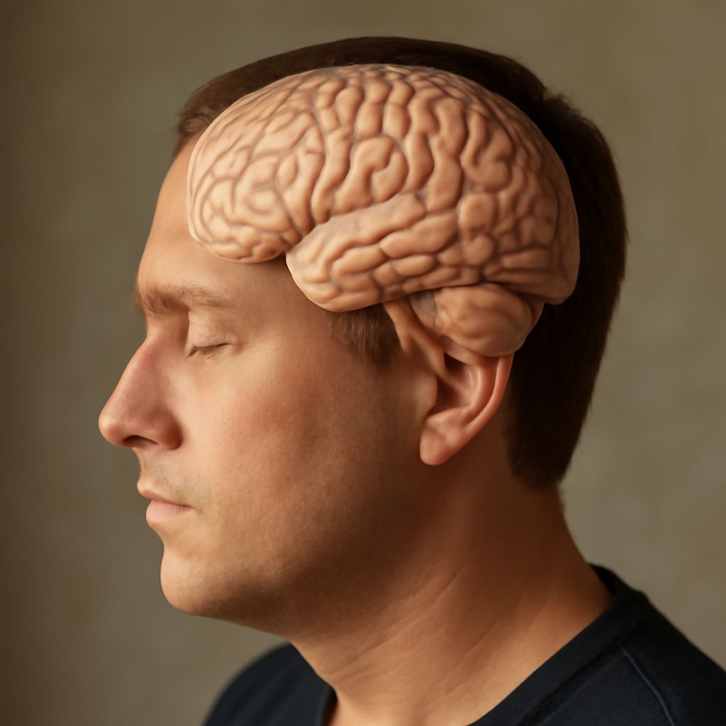
A sheet of tissue only a few millimeters thick folds across the surface of each cerebral hemisphere. Its ridges and grooves make room for a remarkably large area within the skull. To understand what the cortex of the brain is, it helps to begin with that physical sheet: a living layer involved in seeing, moving, remembering, and making sense of the world.
This outer layer is part of a much larger nervous system. Its regions have different connections and different specialties, but a familiar act such as reading a sentence depends on their cooperation. A map of the lobes is therefore a useful introduction, provided that the map is not mistaken for a collection of independent mental compartments.
What Is the Cortex of the Brain? The anatomy beneath the folds
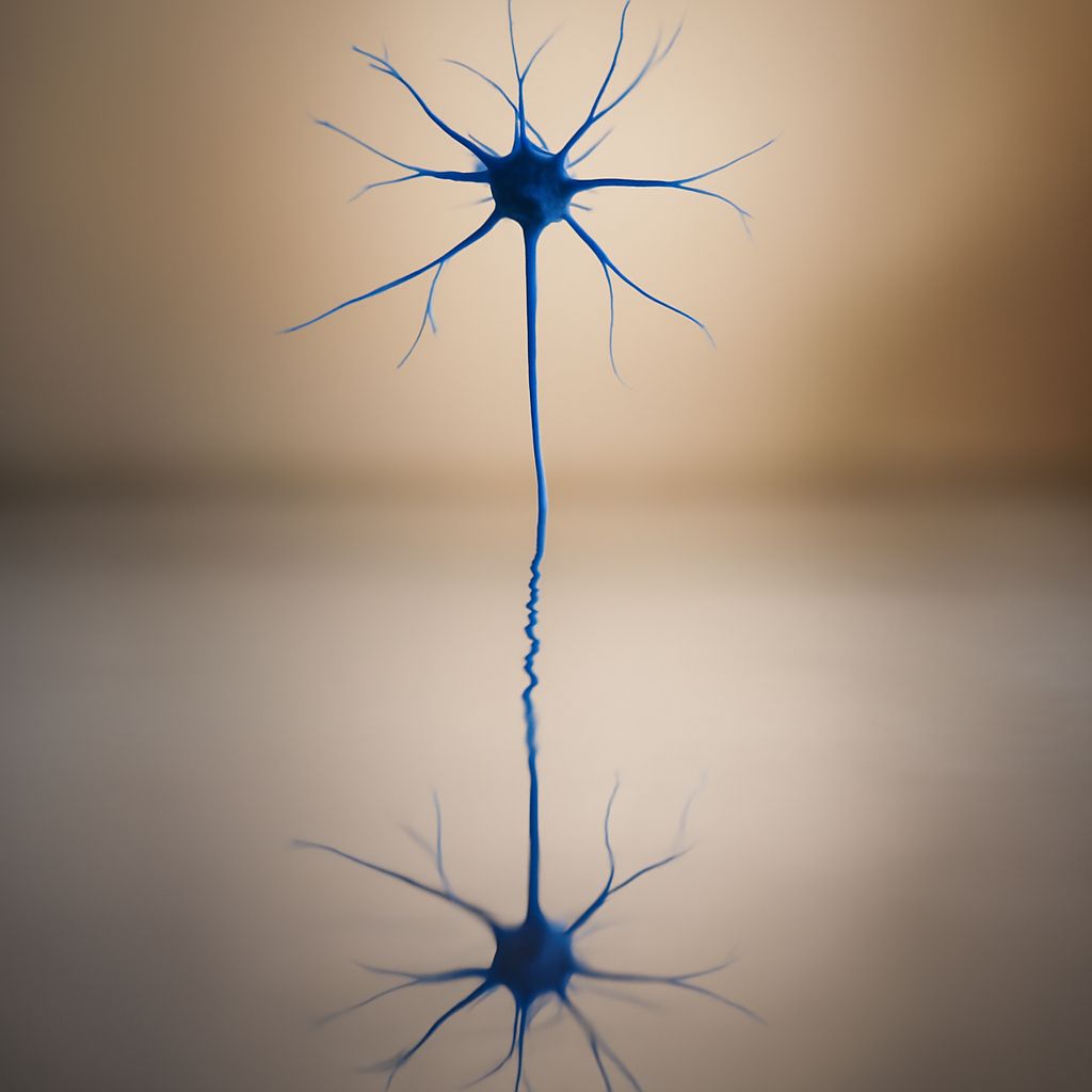
What is the cortex of the brain? It is the outer layer of gray matter covering the cerebral hemispheres, the paired halves of the cerebrum. Gray matter contains nerve cell bodies, dendrites, synapses, and supporting cells. Beneath much of it lies white matter, which contains bundles of nerve fibers connecting cortical regions with one another and with structures deeper in the nervous system. The OpenStax anatomy text describes how the folds increase the available cortical surface area.
The raised folds are called gyri; the grooves between them are sulci. Some prominent grooves help anatomists identify boundaries, although no two individuals have precisely the same pattern. A deep longitudinal fissure separates the hemispheres, and the corpus callosum carries many connections between them. Each hemisphere has an outer surface, an inner surface facing the other hemisphere, and an underside; substantial areas are hidden in folds or along those surfaces.
Much of the brain cortex is neocortex, organized in six layers of cells. The layers differ in the cells they contain and the connections they make. Other cortical regions have a different organization: parts of the hippocampal formation, for example, have fewer layers. This distinction helps explain what the brain’s cortex is: an anatomical arrangement rather than a single uniform circuit. The Duke University Medical School anatomy guide gives an overview of these variations and the underlying pathways.
Four lobes, many kinds of work
The most familiar map divides each hemisphere into frontal, parietal, temporal, and occipital lobes. These labels identify broad territories, not single-purpose organs. The insula is another cortical region, buried deep within the lateral sulcus, beneath parts of the frontal, parietal, and temporal lobes. Anatomical descriptions may also group regions on the inner surface into a limbic lobe. Such labels capture different features of a complex landscape.
Frontal lobes lie toward the front of the head. They include the primary motor cortex, just ahead of the central sulcus, and areas involved in planning and coordinating actions. Other frontal regions contribute to holding information in mind, directing attention, and evaluating possible actions. Speaking also recruits frontal areas, including a region traditionally called Broca’s area, alongside a wider language network.
Parietal lobes lie behind the central sulcus and contribute to processing information about the body and its position in space. The primary somatosensory cortex, just behind that groove, receives information related to touch and other bodily sensations. Other parietal regions help combine sensory information and guide attention toward relevant locations. Reaching for a cup, for instance, calls on information about both the hand and the cup’s position.
Temporal lobes sit roughly at the sides of the cerebrum. Parts of their upper surface are important for processing sound; other areas participate in understanding language and recognizing complex visual patterns. Structures in the medial temporal region, including the hippocampal formation, are important for forming certain kinds of memory. A voice can be heard as sound and recognized as belonging to a particular person through contributions from several connected areas.
Occipital lobes, at the back of the cerebrum, contain primary visual cortex and other visual areas. Signals from the eyes reach visual cortex through pathways that include the thalamus. Subsequent processing involves multiple regions, including areas beyond the occipital lobes, as the nervous system extracts features and relates them to action and recognition. The OpenStax overview provides a starting point for the lobes and their broad roles.
How connections turn a map into a working system
Brain cortex function depends on communication across regions. The functions of this cerebral layer emerge through these connections. Consider the ordinary task of listening to a friend in a crowded room: auditory areas process aspects of sound, language-related regions contribute to meaning, and attention-related networks help select a voice amid competing noise. Memory and expectations can shape interpretation. The exact pattern varies with the task and the person. Attention can shift when another voice calls a name, while the meaning of a sentence may become clear only after its final word. These changes unfold over fractions of a second and longer intervals.
These connections run both within and between hemispheres and also link cortex with the thalamus, basal ganglia, brainstem, and other structures. White-matter pathways provide physical routes for signals; coordinated activity across regions can reveal functional networks. A route and a functional network are related ideas, but a measure of synchronized activity alone does not establish the direction of information flow or prove a direct connection. The NINDS imaging definitions distinguish several types of white-matter pathways.
In the brain, cerebral cortex and subcortical structures participate together in perception and action. The thalamus, for example, relays and regulates information in circuits with the cortex. The cerebellum also contributes to coordination through connections with other brain systems. Describing a cortical region’s contribution is therefore more precise than assigning an entire experience to one spot.
Specialization without a one-to-one code
The outer layer contains regions whose organization reflects particular inputs and outputs. Neighboring points on parts of the primary somatosensory cortex represent neighboring areas of the body, although the amount of cortex devoted to a body part is not simply proportional to its physical size. The hands and face have relatively extensive representations, reflecting the importance of fine sensory distinctions there.
Specialization also appears across the two hemispheres. Language functions are often more strongly represented in the left hemisphere, but the degree and pattern vary between people, and the right hemisphere contributes to aspects of communication. Both hemispheres ordinarily work together. A lobe’s contribution to an activity is thus a question of connections and circumstances, not a license to sort people into fixed “left-brained” and “right-brained” types.
Researchers investigate these relationships with complementary methods. Imaging can associate patterns of activity with a task; studies of anatomy trace pathways; observations of changes after injury can show what a region contributes. Each method has limits. Activity in an area during a task does not, on its own, show that the area performs the entire task or is uniquely necessary for it.
A familiar activity, seen differently
Reading a note brings the organization into focus. Visual pathways carry information about marks on the page; cortical networks contribute to recognizing words and relating them to language and memory. If the reader reaches for a pen to reply, sensory and motor systems participate in guiding the hand. The sequence feels continuous because many processes are coordinated over time.
Understanding what the brain’s cortex is means seeing a folded surface with recognizable regions and connections among them. Following a sentence from the page to an intended response makes that architecture tangible: a familiar moment draws on specialized parts of a distributed system working together.